Detecting Glaucoma Through AI: Advancing Early Diagnosis and Preventing Vision Loss
Leveraging Artificial Intelligence for Automated Optic Nerve Analysis, Early Risk Identification, and Scalable Retinal Screening
Key Highlights
- AI-powered calculation of Cup-to-Disc Ratio (CDR) from a single fundus image
- Automated optic disc and optic cup segmentation for glaucoma risk assessment
- Supports early identification of glaucoma suspects for timely referral
- Reduces manual assessment time and variability in interpretation
- Enables rapid, scalable, and accessible retinal screening
- Designed to support clinicians through AI-assisted retinal analysis
- Expanding AI development pipeline for additional retinal diseases and abnormalities
Introduction
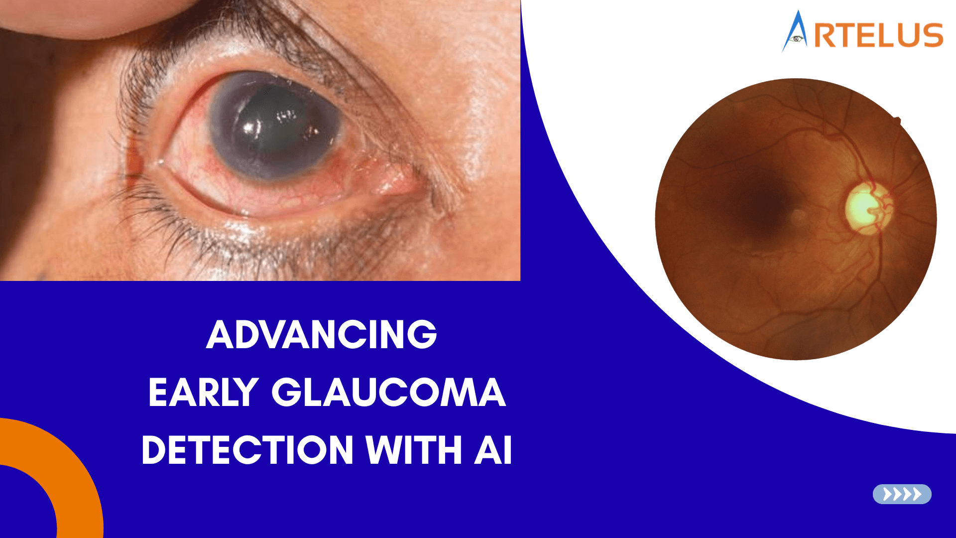
Glaucoma is one of the leading causes of irreversible blindness worldwide and is often referred to as the "silent thief of sight." The disease can progress gradually without noticeable symptoms, causing permanent damage to the optic nerve before a patient becomes aware of any vision changes.
Early detection is critical because vision loss caused by glaucoma cannot be reversed. However, access to routine eye examinations and specialist care remains limited for many populations, particularly in underserved and remote regions.
Advances in Artificial Intelligence (AI) are creating new opportunities to identify glaucoma risk earlier and more efficiently. By analyzing retinal images, AI can assist clinicians in detecting subtle structural changes associated with glaucoma, supporting timely intervention and better patient outcomes.
Understanding Glaucoma
Glaucoma is a group of eye diseases characterized by damage to the optic nerve, which plays a vital role in transmitting visual information from the eye to the brain.
If left undetected and untreated, glaucoma can progressively reduce peripheral vision and eventually lead to irreversible blindness. Since the early stages often present no symptoms, many individuals remain undiagnosed until significant vision loss has already occurred.
This makes routine screening and early diagnosis essential components of preventive eye care.
The Role of the Cup-to-Disc Ratio (CDR)
One of the most important indicators used in glaucoma assessment is the Cup-to-Disc Ratio (CDR).
The optic disc is the region of the retina where the optic nerve exits the eye. At its center lies a depression known as the optic cup.
In healthy eyes, the optic cup occupies a relatively small portion of the optic disc. As glaucoma progresses and optic nerve fibers are damaged, the cup enlarges, increasing the Cup-to-Disc Ratio.
A higher-than-normal CDR may indicate optic nerve damage and can serve as an important marker for glaucoma risk.
Traditionally, ophthalmologists assess CDR through retinal examination and image interpretation. However, this process can be time-consuming and may vary between observers.
How AI Supports Glaucoma Detection
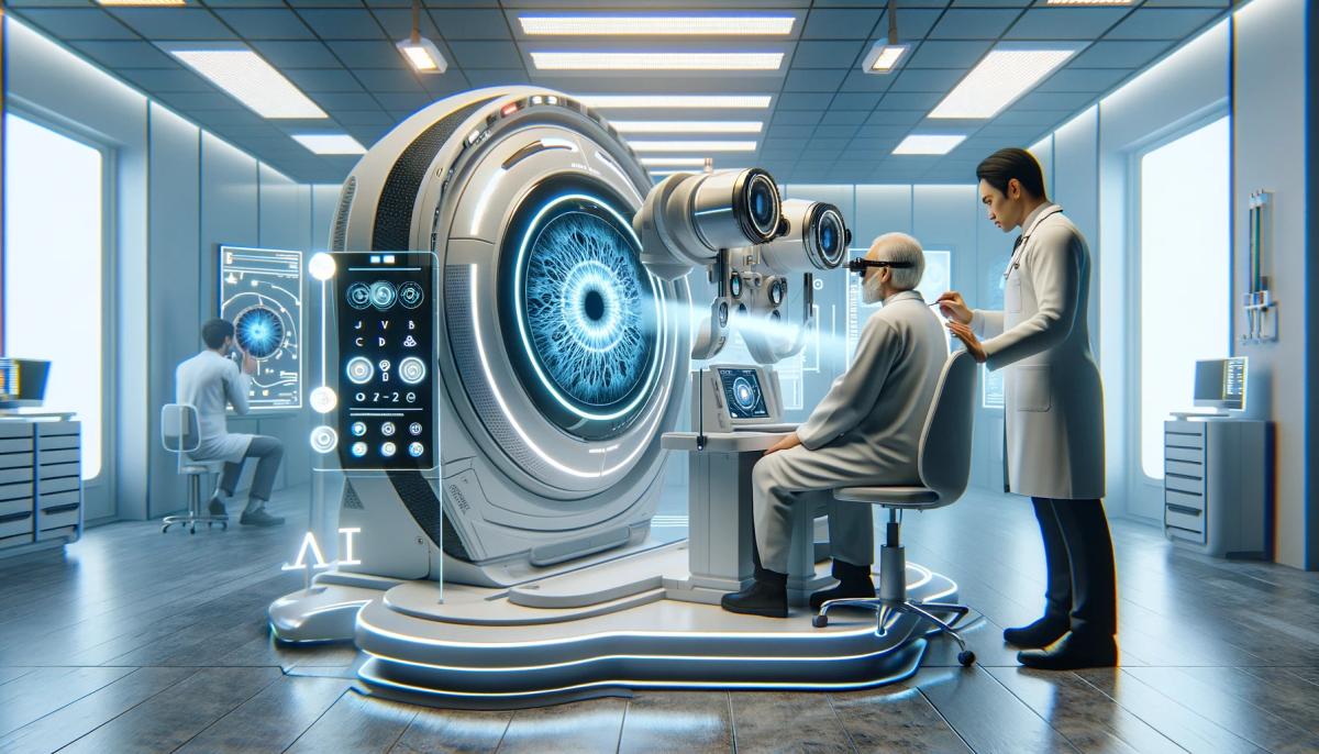
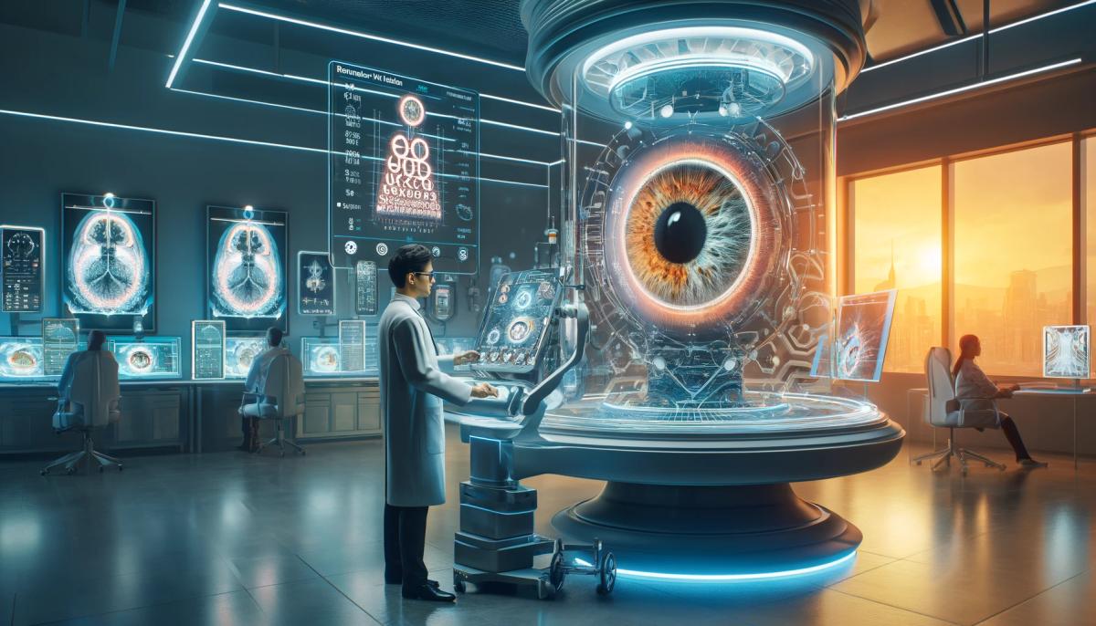
Artificial Intelligence can enhance glaucoma screening by automatically analyzing retinal fundus images and identifying structural changes associated with optic nerve damage.
Using advanced image processing and deep learning techniques, AI systems can:
- Detect and segment the optic disc
- Identify the optic cup
- Calculate the Cup-to-Disc Ratio
- Flag potential glaucoma suspects for further evaluation
- Support clinicians with objective measurements and insights
This enables faster and more consistent screening while reducing the burden of manual assessment.
Artelus AI for Glaucoma Risk Assessment
Artelus has developed AI systems capable of calculating the Cup-to-Disc Ratio from a single fundus image.
Using AI-driven optic disc and optic cup segmentation, the system automatically analyzes retinal images and generates objective measurements that can support glaucoma risk assessment.
When the calculated CDR exceeds clinically relevant thresholds, patients may be identified as glaucoma suspects and referred for comprehensive ophthalmic evaluation.
Key benefits include:
- Automated CDR calculation
- Rapid image analysis
- Reduced manual interpretation time
- Consistent and repeatable measurements
- Scalable screening capabilities
- Improved access to early risk identification
By combining retinal imaging with intelligent analysis, Artelus aims to support healthcare professionals in delivering timely and efficient eye care.
Expanding AI Beyond Glaucoma Detection
Artelus continues to invest in the development of advanced retinal AI technologies designed to support broader retinal disease detection and screening.
In addition to glaucoma risk assessment, AI capabilities currently under development include:
- Glaucoma Risk Assessment
- Age-related Macular Degeneration (AMD / ARMD)
- Drusen
- Central Serous Retinopathy (CSR)
- Choroidal Neovascularization (CNV)
- Macular Hole
- Other retinal abnormalities
These developments are intended to support more comprehensive retinal screening workflows and help clinicians identify a wider range of vision-threatening conditions at earlier stages.
By expanding AI-powered retinal analysis, Artelus is working toward making advanced eye care more accessible, scalable, and impactful across diverse healthcare environments.
Expanding Access to Preventive Eye Care
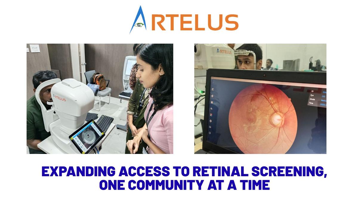
One of the greatest advantages of AI-powered retinal analysis is its ability to extend screening services beyond specialized eye care centers.
With appropriate retinal imaging systems, AI-assisted screening can support:
- Hospitals and eye clinics
- Community health programs
- Vision screening camps
- Primary healthcare centers
- Rural and remote healthcare facilities
This approach can help identify at-risk individuals earlier, strengthen referral pathways, and improve access to preventive eye care services.
For populations that traditionally face barriers to specialist care, AI has the potential to bridge critical gaps in screening and diagnosis.
The Future of AI in Retinal Diagnostics
Artificial Intelligence is rapidly transforming ophthalmology by enabling faster, more scalable, and data-driven approaches to retinal disease detection.
Future advancements may support:
- Multi-disease retinal screening
- Longitudinal disease monitoring
- Predictive risk assessment
- Clinical decision support
- Population-scale screening programs
- Improved healthcare accessibility
As these technologies continue to evolve, AI will play an increasingly important role in helping clinicians detect disease earlier and preserve vision for more patients worldwide.
Conclusion
Glaucoma remains a major cause of preventable vision loss, but early detection can significantly improve outcomes.
AI-powered retinal analysis offers a promising approach to glaucoma risk assessment by enabling automated Cup-to-Disc Ratio measurement, rapid image interpretation, and scalable screening.
Through continued innovation in retinal imaging and artificial intelligence, Artelus is working toward a future where advanced eye care technologies support earlier diagnosis, timely intervention, and broader access to vision-preserving healthcare.
Learn More About Artelus
Artelus is committed to advancing retinal diagnostics through AI-powered technologies designed to improve accessibility, efficiency, and early disease detection.
To learn more about our retinal imaging and AI solutions, or to explore partnership opportunities, contact us at:
📧 contact@artelus.com
Your Vision. Our Mission. Get Screened today with Artelus.