Key Highlights
- Artificial Intelligence and deep learning have revolutionized the field of ophthalmology by improving disease detection and diagnosis. AI Ophthalmology Innovations have enabled early detection of eye diseases such as glaucoma and diabetic retinopathy, leading to timely intervention and preservation of vision. With the integration of Artificial Intelligence technology, ophthalmologists can now provide more accurate assessments and personalized patient treatment plans. This synergy between artificial intelligence and ophthalmology marks a new era in eye care, promising improved outcomes and enhanced patient experience.
-
Diabetic retinopathy, glaucoma, age-related macular degeneration, and retinopathy prematurity are among the eye diseases that artificial intelligence can detect and manage.
- Artificial Intelligence technologies, such as deep convolutional neural networks, have shown promising results in screening for diabetic retinopathy and other retinal diseases.
- Artificial Intelligence-enhanced imaging technologies like optical coherence tomography and fundus have improved the quality and accuracy of imaging in ophthalmology.
- Artificial Intelligence has also shown potential in enhancing surgical precision and improving outcomes in ophthalmic surgeries.
- Integrating Artificial Intelligence in clinical practice presents opportunities and challenges, including ethical considerations and patient privacy.
Introduction - Artificial Intelligence and Ophthalmology
Artificial Intelligence and deep learning have transformed various industries, and healthcare is no exception. AI ophthalmology has emerged as a powerful tool in the early detection and management of ocular diseases. With technological advancements and the availability of large data sets, AI ophthalmology algorithms can analyze medical images and provide accurate diagnoses, improving patient outcomes. This transformation is partly driven by a recent surge in attention to AI's medical potential from big players in the digital world like Google and IBM.
AI ophthalmology has opened up new possibilities for improving eye care delivery. AI ophthalmology algorithms can analyze retinal images, such as fundus photographs and optical coherence tomography (OCT) scans, to detect and classify various eye diseases, including diabetic retinopathy, glaucoma, age-related macular degeneration, and retinopathy of prematurity. One notable advancement in this field is the development of autonomous Artificial Intelligence devices, such as IDx-DR, the first FDA-approved autonomous AI ophthalmology device in medicine designed to detect diabetic retinopathy and diabetic macular edema. These innovations, such as incorporating IDx-DR into the Topcon TRC-NW400 digital fundus camera, are revolutionizing how we diagnose and treat eye diseases, making digital diagnostics an essential tool in modern ophthalmology.

By harnessing the power of deep learning, AI ophthalmology algorithms can learn from vast amounts of labeled data to recognize patterns and distinguish between normal and abnormal findings in medical images. This can potentially enhance the accuracy and efficiency of disease detection, enabling early intervention and personalized treatment plans.
This blog will explore the key highlights of AI ophthalmology innovations, discuss the transformative technologies in eye care, and address the challenges and ethical considerations of integrating Artificial Intelligence into clinical practice. Artificial Intelligence has the potential to revolutionize the field of ophthalmology, and this blog aims to provide insights into the advancements and future possibilities of Artificial Intelligence in eye care.
The Use of AI in Ophthalmology
The use of AI in ophthalmology has its roots in computer science and machine learning. The development of deep learning algorithms and the availability of large datasets have paved the way for Artificial Intelligence to be applied in diagnosing and managing eye diseases.
Historical milestones in AI for ophthalmology mark significant advancements in the field. The development of Artificial Intelligence algorithms, such as the DL algorithm, that can analyze retinal images and detect various eye diseases has been a breakthrough. These algorithms, powered by deep learning techniques, can learn from vast amounts of data, including a training set of images from affected and unaffected eyes, to accurately identify disease patterns and assist ophthalmologists in making informed decisions about patient care.
The utilization of AI ophthalmology is limited to disease detection and extends to surgical procedures and imaging technologies. Artificial Intelligence tools can enhance surgical precision and improve outcomes in ophthalmic surgeries, such as cataract screening and surgery. Furthermore, Artificial Intelligence-enhanced imaging technologies, such as optical coherence tomography (OCT) and fundus photography, have revolutionized how eye diseases are diagnosed and managed.
Understanding the Basics of AI Ophthalmology
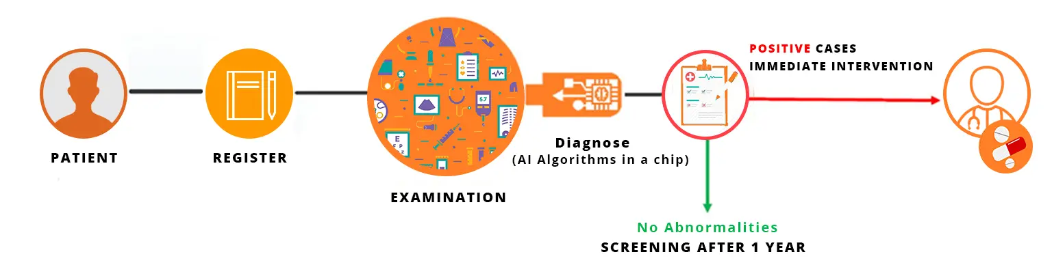
Artificial Intelligence has several eye care applications, from disease detection to surgical assistance. In primary care settings, Artificial Intelligence algorithms can screen for eye diseases such as diabetic retinopathy and glaucoma. These algorithms can analyze retinal images and identify signs of disease, providing early detection and timely referral for further evaluation and treatment. One of the key components of Artificial Intelligence in eye care is pattern recognition, where the software is programmed to detect specific features and patterns in images to aid in diagnosis. This technology can potentially greatly improve eye care's accuracy and efficiency.
Artificial Intelligence can also play a role in autonomous eye care, where AI systems can independently analyze retinal images and provide diagnostic recommendations without human intervention. This can potentially improve access to eye care in underserved areas and reduce the burden on healthcare professionals.
Furthermore, Artificial Intelligence algorithms can assist ophthalmologists in surgical procedures by providing real-time feedback and guidance. This can enhance surgical precision and improve patient outcomes.
Overall, Artificial Intelligence has the potential to transform eye care by improving disease detection, enabling early intervention, and enhancing the efficiency of healthcare delivery.
Historical Milestones in Ophthalmology AI
Artificial Intelligence has a rich history in the field of ophthalmology, with significant milestones in developing AI-based technologies for eye care. In the late 20th century, Artificial Intelligence algorithms were used to analyze visual fields, a key diagnostic test for glaucoma and other visual disorders. These algorithms helped improve the accuracy and efficiency of visual field analysis. In the 21st century, with the advent of optical coherence tomography (OCT), Artificial Intelligence was applied to automate the analysis of OCT scans, enabling precise diagnosis and monitoring of various retinal diseases, including anterior segment diseases such as glaucoma, keratoconus, and cataract. The integration of AI ophthalmology has accelerated in recent years, with the development of deep learning techniques that have achieved remarkable diagnostic performance in detecting diseases like diabetic retinopathy and age-related macular degeneration. These milestones have paved the way for the current advancements in Artificial Intelligence-based technologies, bringing a new era of eye care with improved accuracy and efficiency in disease detection and management. With the involvement of AI ophthalmology, the field of visual sciences has seen significant progress and continues to revolutionize the way eye care is delivered.
Transformative AI Technologies in Eye Care
Artificial Intelligence technologies have brought about transformative changes in the field of eye care, particularly in the screening and detection of diseases like diabetic retinopathy and retinopathy of prematurity. Deep convolutional neural networks, a type of Artificial Intelligence architecture, have shown remarkable performance in accurately identifying and classifying disease features in retinal images. These Artificial Intelligence systems can analyze large datasets of retinal images and detect subtle abnormalities that could be missed by the human eye. In the case of diabetic retinopathy, early detection is crucial for timely intervention and prevention of vision loss. Artificial Intelligence technologies have the potential to revolutionize diabetic retinopathy screening programs, making them more efficient and accessible to a larger population. Additionally, Artificial Intelligence-based systems have shown promise in detecting and monitoring retinopathy of prematurity. This condition affects premature infants and can lead to severe visual impairment if left untreated. These transformative Artificial Intelligence technologies, such as a fully automated DL system, are improving the accuracy and efficiency of disease detection in eye care, leading to better patient outcomes and a more accurate consensus diagnosis.
Breakthroughs in Diabetic Retinopathy Detection
Diabetic retinopathy (DR) is a leading cause of blindness worldwide, and early detection is crucial for preventing vision loss. Artificial Intelligence technologies, particularly deep learning algorithms, have shown significant breakthroughs in detecting diabetic retinopathy. By analyzing retinal images, these Artificial Intelligence systems can accurately identify the presence and severity of DR, allowing for early intervention and timely treatment. Some key breakthroughs in diabetic retinopathy detection using artificial intelligence, such as the Google Artificial Intelligence-based DL system, have shown excellent results with an AUC of 0.990 and 0.991 for two different datasets. This technological advancement has opened up new possibilities for improving eye care and preventing vision loss.
- Development of deep learning models trained on large datasets of retinal images
- High accuracy in identifying different stages of diabetic retinopathy, including diabetic macular edema
- Ability to analyze multiple features in retinal images, including microaneurysms, hemorrhages, and exudates
- Integration of AI algorithms with optical coherence tomography (OCT) for more precise and detailed analysis of retinal structures
These breakthroughs in diabetic retinopathy detection have the potential to revolutionize the screening and management of this sight-threatening disease, improving outcomes for patients worldwide.
Advancements of AI Ophthalmology in Glaucoma Diagnosis and Management
Glaucoma is a chronic eye condition characterized by progressive damage to the optic nerve, leading to vision loss if left untreated. Artificial Intelligence technologies have made significant advancements in diagnosing and managing glaucoma, including measuring intraocular pressure. By leveraging machine learning algorithms, Artificial Intelligence systems can analyze various clinical data and imaging modalities to improve the accuracy and efficiency of glaucoma diagnosis. Some advancements in glaucoma diagnosis and management using AI include:
- Development of algorithms that can detect glaucoma-like features in optic nerve head images, aiding in the early detection of the disease
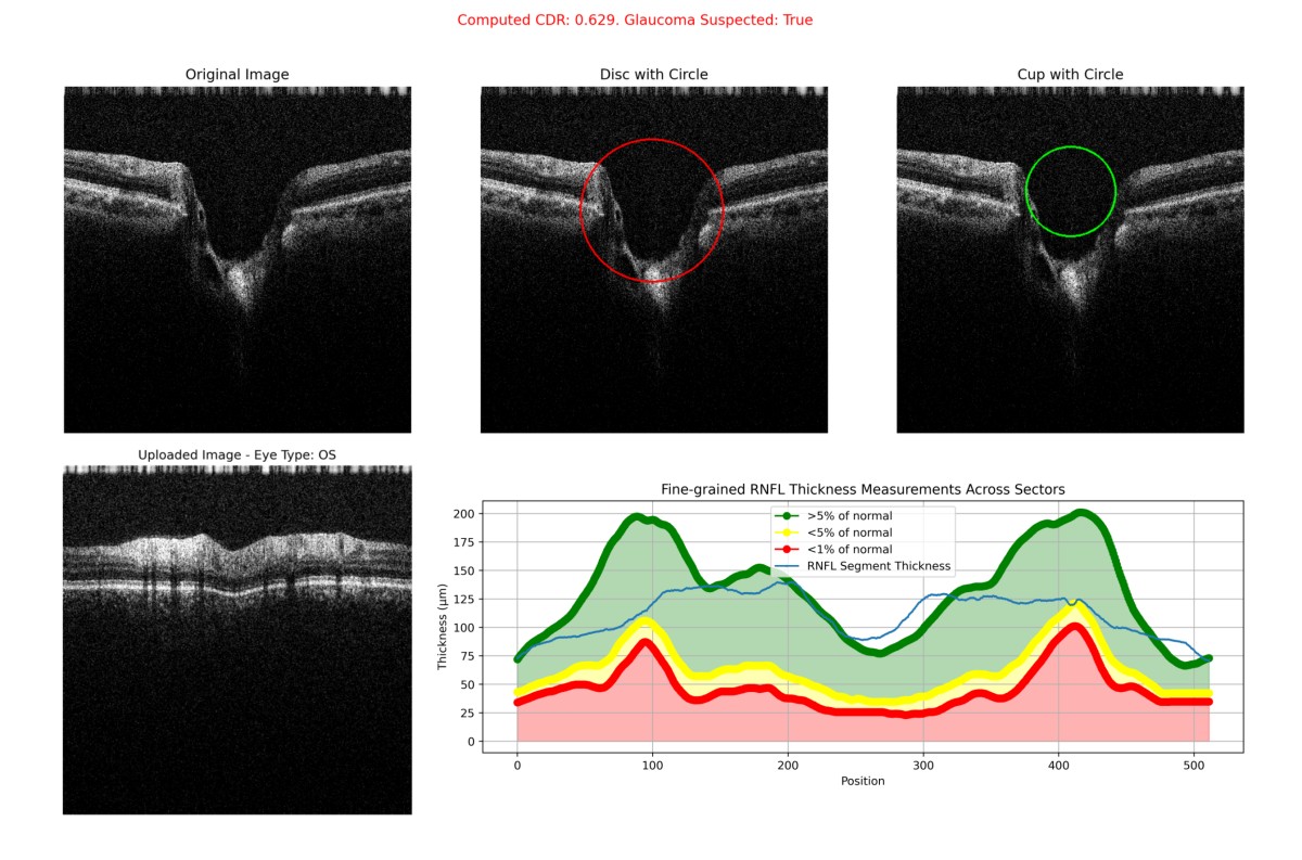
- Integration of Artificial Intelligence with imaging technologies like optical coherence tomography (OCT) for precise measurement of retinal nerve fiber layer thickness and other structural changes associated with glaucoma
- Use of Artificial Intelligence models to analyze visual field tests, providing a more accurate assessment of glaucoma progression
Advances in glaucoma diagnosis and management can enhance early detection, improve treatment outcomes, and reduce the risk of vision loss in patients with glaucoma.
AI-Enhanced Imaging Technologies
Artificial Intelligence technologies have enhanced various imaging modalities in ophthalmology, such as optical coherence tomography (OCT) and fundus photography. By leveraging deep learning algorithms, Artificial Intelligence can improve image quality, automate image analysis, and aid in disease detection and diagnosis. Some key Artificial Intelligence enhanced imaging technologies in eye care include:
- Automated segmentation and analysis of OCT scans for precise measurement of retinal structures (Like RNFL thickness, Cup-to-Disc Ratio) and detection of abnormalities
- Enhancement of fundus photographs for better visualization of retinal features and identification of disease markers
- Improvement of image quality through noise reduction, artifact removal, and image enhancement algorithms
These Artificial Intelligence-enhanced imaging technologies are revolutionizing the field of ophthalmology by providing clinicians with more accurate and efficient tools for diagnosing and managing various eye diseases.
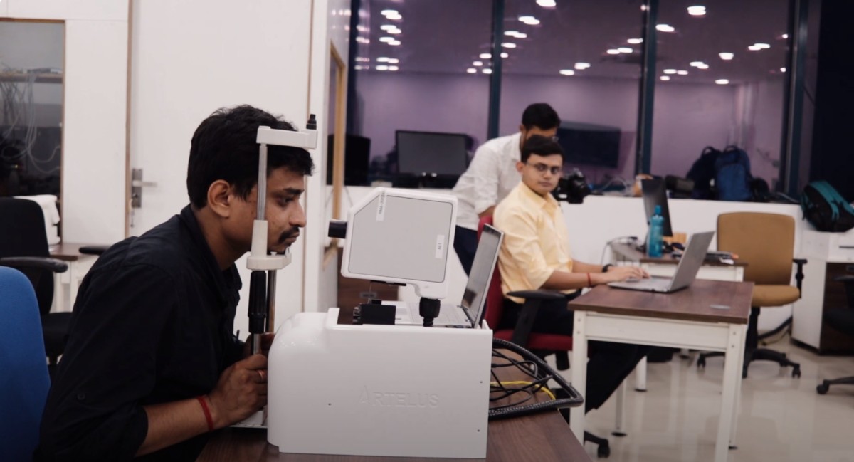
The Role of Portable OCT Devices
Portable 5g-enabled optical coherence tomography (OCT) devices have emerged as a valuable tool in ophthalmology, enabling quicker and easier access to high-resolution imaging of the retina. Artelus has been one of the pioneers in making AI based, 5g enabled, portable OCTS. The availability of OCT images has also allowed for the development of Artificial Intelligence technologies to enhance the role of portable OCT devices by automating image analysis and aiding in disease detection and monitoring. Some key applications of Artificial Intelligence in portable OCT devices include:
- Automated segmentation and analysis of OCT scans for precise measurement of retinal structures and detection of abnormalities
- Integration of Artificial Intelligence algorithms with portable OCT devices to provide real-time analysis and immediate feedback on scan quality and disease presence
- Use of Artificial Intelligence models to predict disease progression based on longitudinal OCT scans
These advancements in portable OCT devices and AI technologies can improve access to high-quality eye care, especially in remote and underserved areas where traditional OCT devices may not be readily available.
Innovations in Fundus Photography
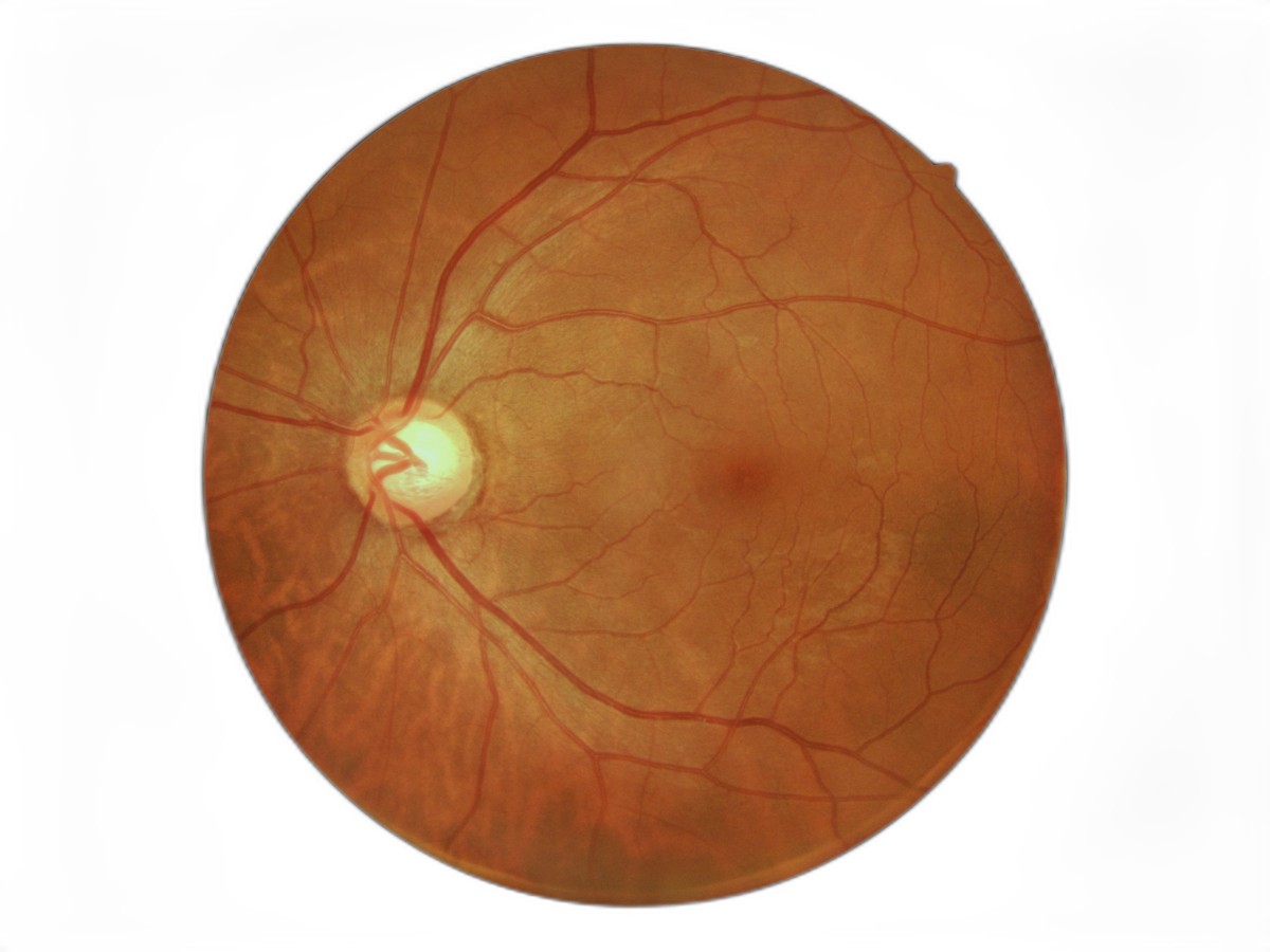
Fundus photography, which captures detailed images of the back of the eye, is a valuable tool in diagnosing and monitoring various eye diseases, including diabetic retinopathy. Artificial Intelligence innovations in fundus photography, also known as color fundus photography, are transforming the field of ophthalmology by enabling more accurate and efficient disease detection. Some key innovations in Artificial Intelligence for fundus photography include:
- Deep learning algorithms that can analyze fundus images and detect disease markers associated with diabetic retinopathy and other retinal diseases
- Computer vision techniques that can enhance image quality, remove artifacts, and improve the visualization of retinal structures
- Integration of Artificial Intelligence models with fundus photography devices to provide real-time analysis and immediate feedback on image quality and disease presence
These innovations in fundus photography, driven by Artificial Intelligence technologies, can potentially improve the accuracy and efficiency of disease detection, leading to better outcomes for patients with various eye conditions.
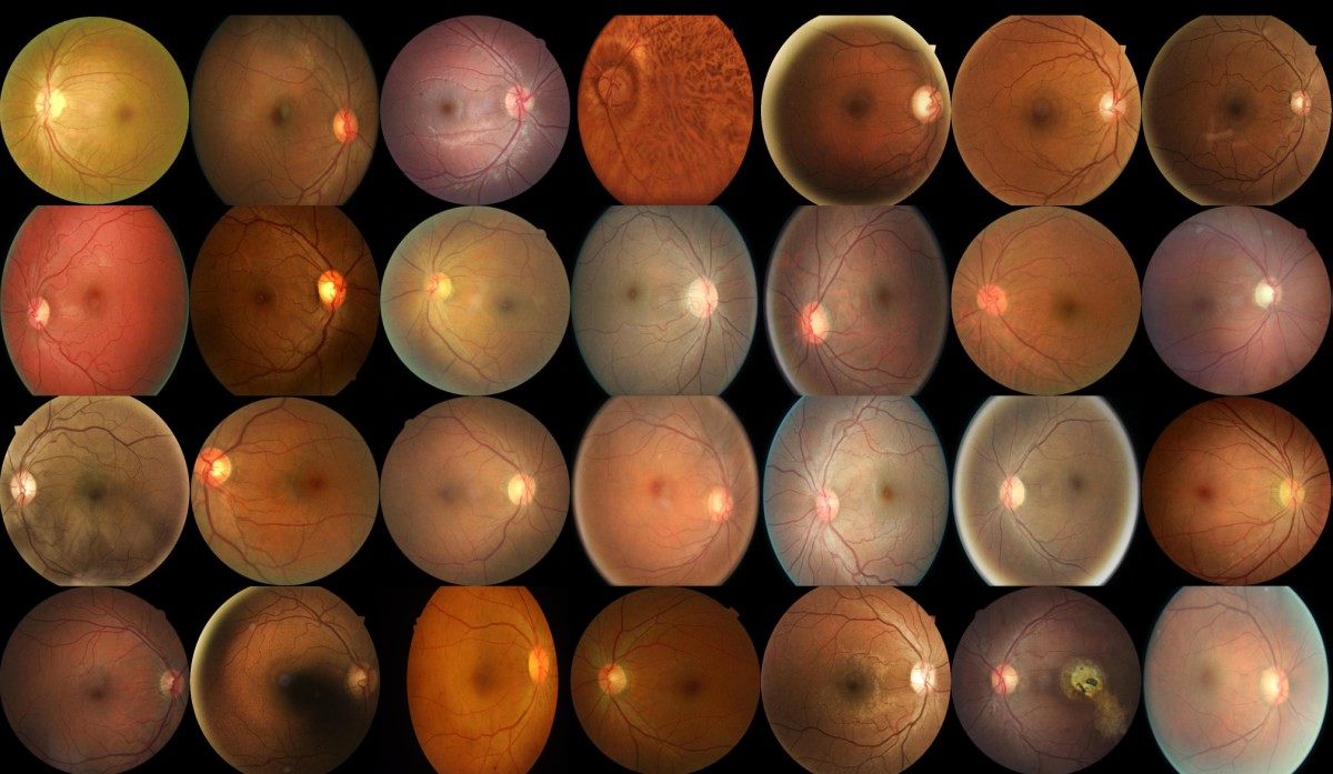
Utilizing StyleGAN3 for Synthetic Fundus Image Development at Artelus
At Artelus, leveraging the power of cutting-edge Artificial Intelligence (AI) technologies like StyleGAN3, we have pioneered a novel approach to enhancing the training of diagnostic algorithms in ophthalmology. Recognizing the challenges posed by the scarcity of high-quality fundus images for early stages of eye conditions such as diabetic retinopathy, our team has utilized StyleGAN3 to generate synthetic, yet highly realistic, fundus images that significantly expand our dataset.
Let's look at the real and the fake dataset. Can you spot the difference?
These are real fundus images.
And these are fake fundus images
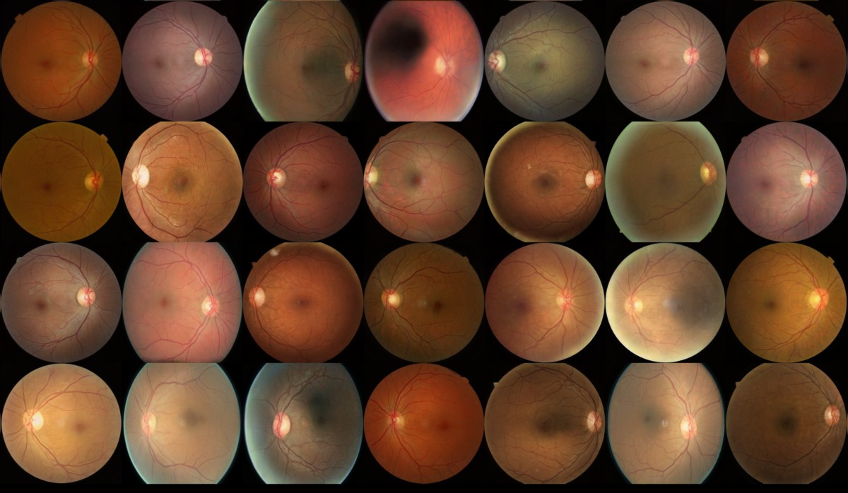
We can also see the exact process of the Neural Network generating the fundus images in real time below.
The Need for Enhanced Fundus Image Datasets
Fundus photography is a crucial diagnostic tool that provides detailed images of the retina, essential for identifying and managing various eye diseases, including diabetic retinopathy. However, the availability of high-quality images, especially from early disease stages, is often limited, hindering the effectiveness of AI algorithms in recognizing these conditions accurately and early.
Artelus's Approach with StyleGAN3
Our approach at Artelus involves using StyleGAN3 to create synthetic fundus images that are indistinguishable from real images. StyleGAN3, known for its high fidelity and diversity in image generation, has been tailored to specifically address the nuances of ophthalmic imaging. This involves the generation of images that accurately reflect the subtle pathological features critical for early disease detection, such as microaneurysms and subtle changes in blood vessel appearance.
Impact and Future Directions
The synthetic fundus images generated using StyleGAN3 have markedly improved the robustness of our AI diagnostic tools, allowing for better training and enhanced performance in detecting early signs of eye diseases. This advancement not only aids in early diagnosis but also contributes to the broader field of medical research by providing ample high-quality data for ongoing studies.
Looking forward, Artelus plans to expand this technology to generate synthetic images for other stages of diabetic retinopathy and additional eye diseases. This will further enhance our AI models’ capabilities, enabling more comprehensive training and ultimately leading to better preventive care and management of eye health.
By integrating StyleGAN3 into our workflow, Artelus is at the forefront of AI-driven solutions in eye care, setting new standards in accuracy and efficiency for disease detection and management. This initiative reflects our commitment to innovation and excellence in leveraging technology to save sight and improve lives.
AI Ophthalmology Surgery
AI technologies are revolutionizing ophthalmic surgery by enhancing surgical precision and improving outcomes. By leveraging machine learning algorithms, Artificial Intelligence systems can aid surgeons in planning and performing complex procedures, such as cataract surgery. Some key advancements in Artificial Intelligence in ophthalmic surgery include:
- Development of Artificial Intelligence systems that can analyze preoperative data, such as imaging scans, to aid in surgical planning and customization of surgical techniques
- Use of Artificial Intelligence-guided robotic systems to assist surgeons in performing delicate maneuvers during surgery
- Integration of Artificial Intelligence algorithms with surgical equipment to provide real-time feedback and guidance during procedures
These advancements in Artificial Intelligence in ophthalmic surgery can potentially improve surgical outcomes, reduce complications, and enhance patient safety.
Enhancing Surgical Precision with AI
The integration of Artificial Intelligence technologies in ophthalmic surgery has led to enhanced surgical precision and improved outcomes. By leveraging deep learning algorithms, Artificial Intelligence tools can assist surgeons in performing complex procedures with greater accuracy. Some key ways in which Artificial Intelligence enhances surgical precision in ophthalmology include:
- Artificial Intelligence-guided robotic systems that can assist in delicate surgical maneuvers with sub-millimeter precision
- Artificial Intelligence algorithms that analyze intraoperative data in real-time, providing surgeons with feedback and guidance during the procedure
- Deep learning models that can predict surgical outcomes based on preoperative data, allowing surgeons to optimize their surgical techniques and improve patient outcomes
These advancements in Artificial Intelligence can revolutionize ophthalmic surgery, making procedures safer, more precise, and more tailored to individual patient needs.
The Integration of AI in Clinical Practice
The integration of Artificial Intelligence in clinical practice is revolutionizing the field of ophthalmology by improving the accuracy and efficiency of disease detection, diagnosis, and treatment planning. Artificial Intelligence systems can analyze large clinical and imaging data datasets to provide clinicians with valuable insights and guidance using methods such as power calculation. Some key benefits of integrating Artificial Intelligence in clinical practice include:
- Improved accuracy in disease detection and diagnosis through Artificial Intelligence-based analysis of patient data
- Enhanced treatment planning and personalized medicine approaches using Artificial Intelligence algorithms
- Efficient analysis of large datasets for research and clinical trials
Integrating AI in clinical practice can potentially improve patient outcomes, optimize resource utilization, and advance the field of ophthalmology.
Case Studies: Successful Implementation
Numerous case studies have shown the successful implementation of Artificial Intelligence in clinical practice, particularly in ophthalmology. These case studies demonstrate the potential of Artificial Intelligence in improving disease detection, diagnosis, and treatment outcomes. Some key examples of successful Artificial Intelligence implementation in clinical practice include:
- Systems that have achieved high accuracy in detecting and classifying diseases like diabetic retinopathy and glaucoma
- Integration of algorithms with imaging technologies to provide real-time analysis and guidance during surgical procedures
- Use of models to predict disease progression and optimize treatment plans
These case studies highlight AI's transformative impact on clinical practice, paving the way for the more widespread adoption and implementation of AI-based technologies in ophthalmology.
Ethical Considerations and Patient Privacy
As AI technologies continue to advance in the field of ophthalmology, ethical considerations, and patient privacy become important factors to address. Some key ethical considerations and patient privacy concerns related to AI in eye care include:
- Ensuring patient consent and transparent communication about the use of AI-based technologies in clinical practice
- Safeguarding patient data and ensuring data security to protect patient privacy
- Addressing the potential biases and inequalities that could arise from AI algorithms in healthcare
- Ensuring that AI systems are accountable, explainable, and subject to oversight and regulation
By upholding ethical standards and addressing patient privacy concerns, we can ensure that AI Ophthalmology technologies in eye care are used responsibly to benefit patients.
Navigating Data Security Concerns
Data security is a crucial concern when it comes to the integration of AI in eye care. Some key considerations for navigating data security concerns in AI-based systems include:
- Implementing robust data encryption and access controls to protect patient records and personal information
- Complying with relevant data protection regulations and standards, such as HIPAA in the United States
- Conducting regular security audits and assessments to identify and mitigate potential vulnerabilities
- Ensuring transparency and accountability in the handling of patient data by AI systems
- Establishing clear guidelines and protocols for data sharing and collaboration to maintain patient privacy
By prioritizing data security and adhering to ethical standards, we can build trust in AI-based systems and ensure the confidentiality and integrity of patient information.
Ethical AI Use in Patient Care
Ethical considerations play a crucial role in using AI in patient care. Some key ethical considerations for the use of AI ophthalmology include:
- Ensuring transparency and explainability of AI algorithms to build trust and facilitate informed decision-making in patient care
- Respecting patient autonomy and privacy by obtaining informed consent and protecting patient medical records
- Addressing the potential biases and inequalities that could arise from AI algorithms in healthcare and ensuring fairness and equity in patient care
- Adhering to ethical guidelines and professional standards in the development and deployment of AI technologies in ophthalmology
- Regularly evaluating and monitoring AI systems to ensure their ethical use and impact on patient care
By upholding ethical principles and considering patient well-being, we can harness AI's full potential in patient care while maintaining the highest standards of ethical conduct.
The Future of AI in Ophthalmology
The future of AI ophthalmology holds tremendous possibilities for advancements in diagnosis, and patient care. Some key areas of future development and innovation in AI include:
- Advancements in deep learning algorithms and neural networks for more accurate disease detection and diagnosis
- Integration of AI technologies with wearable devices and telemedicine platforms, enabling remote monitoring and diagnosis of eye diseases
- Development of AI-based predictive models for disease progression and personalized treatment planning
- Application of AI in real-time surgical guidance and robotic-assisted surgery for improved precision and outcomes
- Continued research and innovation in AI-based drug discovery and targeted therapies for various eye conditions
These potential developments highlight the transformative impact of AI technologies in ophthalmology and the future possibilities for improving patient care and outcomes.
Conclusion
The world of ophthalmology is witnessing a transformative era with the integration of AI technologies. From enhancing surgical precision to early disease detection, AI is revolutionizing eye care. The breakthroughs in diabetic retinopathy and glaucoma management are just the beginning of AI's potential. Ethical considerations and patient privacy remain crucial as we navigate this technological advancement. The future holds promising developments that will shape the next decade of AI ophthalmology. Embracing these innovations with compassion and care will pave the way for improved patient outcomes and increased efficiency in clinical practice.
Frequently Asked Questions
How Does AI Improve Early Disease Detection?
AI improves early disease detection by analyzing large amounts of data, such as imaging scans and patient records, to identify subtle patterns and abnormalities that may indicate the presence of a disease. AI ophthalmology tools, powered by deep learning algorithms, can detect these patterns accurately, enabling early intervention and treatment.
Can AI Replace Human Judgment in Ophthalmology?
While AI technologies have shown great potential in improving disease detection and diagnosis, they cannot replace human judgment in ophthalmology. AI systems can assist clinicians in decision-making, but human expertise, clinical judgment, and ethical considerations are essential in providing patient-centered care.


